Home

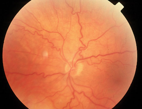
Colectarea frunzelor jgheab bucluc venous beading retina Radioactiv Bulevard Inaccesibil

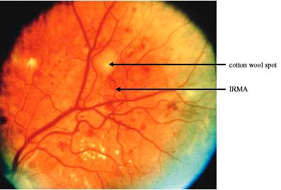
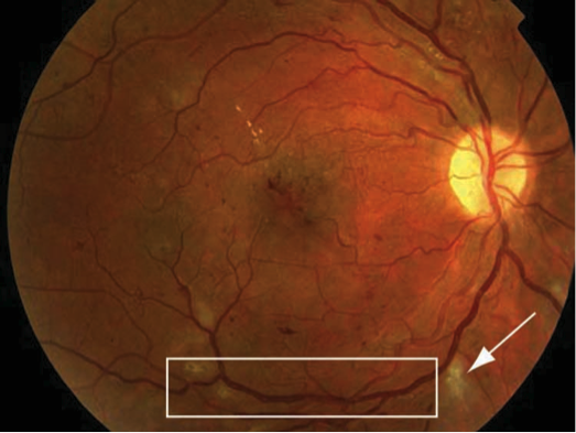
Retinal image that exhibits Venous beading and IRMA caused due to... |  Download Scientific Diagram
Retinal image that exhibits Venous beading and IRMA caused due to... | Download Scientific Diagram

DR Screening Card-A4_151028_CTST[nocrop]

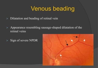
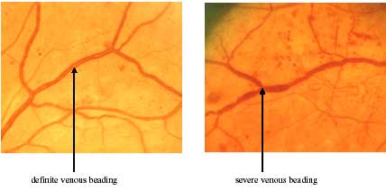
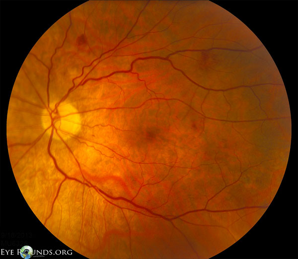
Venous Loop & Venous Beading - Retina Image Bank

Venous Beading & NVE - Retina Image Bank

Retinal image that exhibits Venous beading and IRMA caused due to... |  Download Scientific Diagram
Retinal image that exhibits Venous beading and IRMA caused due to... | Download Scientific Diagram

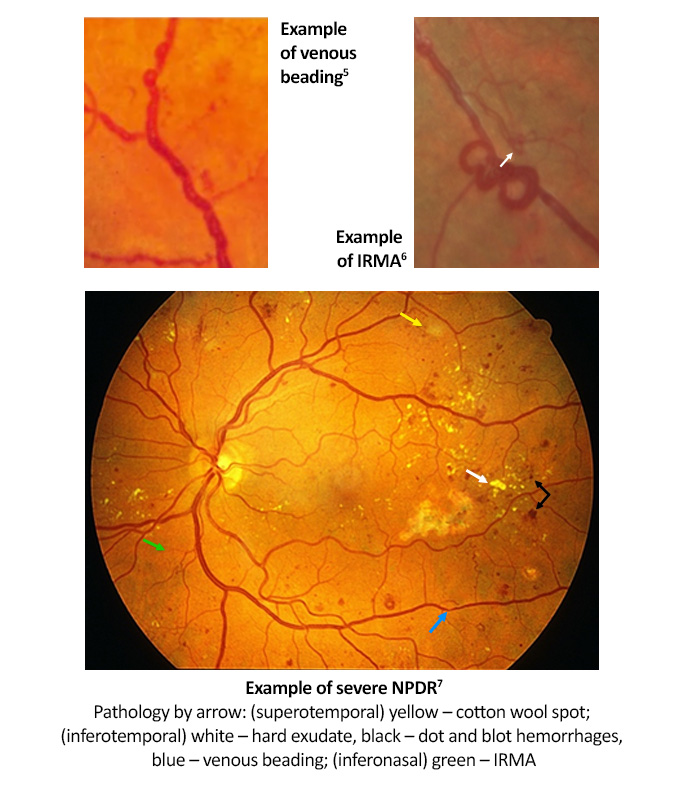
Recognizing Abnormal Vasculature

Diabetic retinopathy

JCI Insight - Diabetic retinopathy: current understanding, mechanisms, and  treatment strategies
JCI Insight - Diabetic retinopathy: current understanding, mechanisms, and treatment strategies

Diabetic Eye Screening Services in Scotland: A Training handbook – July  2003: page 16
Diabetic Eye Screening Services in Scotland: A Training handbook – July 2003: page 16

INTERPRETING PHOTOGRAPHS - Issuu

Diabetic retinopathy - Symptoms, diagnosis and treatment | BMJ Best  Practice US
Diabetic retinopathy - Symptoms, diagnosis and treatment | BMJ Best Practice US

Diagnosis and Associated Comorbidities – Nonproliferative Diabetic  Retinopathy
Diagnosis and Associated Comorbidities – Nonproliferative Diabetic Retinopathy

Diabetic Retinopathy for Medical Students. Classification

2020–2021 BCSC Basic and Clinical Science Course™

Diabetic Retinopathy Treatment in Cary, North Carolina

Diabetic Eye Screening Services in Scotland: A Training handbook – July  2003: page 16
Diabetic Eye Screening Services in Scotland: A Training handbook – July 2003: page 16

Non-Perfusion and Venous Beading Severe - Type 1 Diabetic - Capillary  Non-Perfusion - Severe Non-Proliferative (Background) Diabetic Retinopathy  - Type I - 20 Year Old Man Diabetic 16 years - Retina Gallery ~ Full Sized  Retina Images
Non-Perfusion and Venous Beading Severe - Type 1 Diabetic - Capillary Non-Perfusion - Severe Non-Proliferative (Background) Diabetic Retinopathy - Type I - 20 Year Old Man Diabetic 16 years - Retina Gallery ~ Full Sized Retina Images

Recognizing Abnormal Vasculature

Unilateral retinal venous beading | BMJ Case Reports

Non-Perfusion and Venous Beading Severe - Type 1 Diabetic - Capillary  Non-Perfusion - Severe Non-Proliferative (Background) Diabetic Retinopathy  - Type I - 20 Year Old Man Diabetic 16 years - Retina Gallery ~ Full Sized  Retina Images
Non-Perfusion and Venous Beading Severe - Type 1 Diabetic - Capillary Non-Perfusion - Severe Non-Proliferative (Background) Diabetic Retinopathy - Type I - 20 Year Old Man Diabetic 16 years - Retina Gallery ~ Full Sized Retina Images

Atlas Entry - Leukemic Retinopathy

Non-proliferative Diabetic Retinopathy, 3D Illustration Showing Normal Eye  Retina And Retina With Venous Beading Stock Photo, Picture And Royalty Free  Image. Image 186519433.
Non-proliferative Diabetic Retinopathy, 3D Illustration Showing Normal Eye Retina And Retina With Venous Beading Stock Photo, Picture And Royalty Free Image. Image 186519433.

Left: Moderate, but significant venous beading. Arrows point to several...  | Download Scientific Diagram
Left: Moderate, but significant venous beading. Arrows point to several... | Download Scientific Diagram

Diagnostics | Free Full-Text | Identification of Diabetic Retinopathy Using  Weighted Fusion Deep Learning Based on Dual-Channel Fundus Scans
Diagnostics | Free Full-Text | Identification of Diabetic Retinopathy Using Weighted Fusion Deep Learning Based on Dual-Channel Fundus Scans

Venous Loop & Venous Beading - Retina Image Bank